Opening Hours: Mon - Sat : 6.00 pm - 9.00 pm, Sunday Closed
dr balamurugan
Mail Us Today
shakthieye@gmail.com
Call us for more details
(+91) - 044-23774676 | 63694 70626
Hospital Location
Virugambakkam, Chennai - 600 092
  • Home
  • About Us
  • Services
  • Opticals
  • Disorders of Eye
  • Blogs
  • Testimonial
  • Contact Us
Apppointment

Shakthi Eye Care Centre

Shakthi Eye Care Centre was started in 2018 and ever since its inception it has been offering the most advanced treatments across all areas of eye care. Total eye care as result of years of experience, supported by sincere and ethical patient service to provide the best visual outcome is what that sets it apart!

Read more
Eye clinic near me

Dr. Balamurugan    PR

Dr Balamurugan is an ophthalmologist with more than 20 years of experience in the field of ophthalmology and has done over 20000 cataract surgeries so far. He has undergone training in cornea and external diseases of the eye and has vast experience in diagnosing and treating ocular surface diseases

Read more

About Dr. Balamurugan PR

Dr. Balamurugan PR is an ophthalmologist with more than 20 years of experience in the field of ophthalmology. He completed his MBBS degree in 1994 and MS degree in ophthalmology from Manipal Academy of Higher Education in 1998. He won the Gold Medal for the best outgoing student in MS Ophthalmology from Kasturba Medical college, Manipal Academy of Higher Education.

After his post graduate training, he did a fellowship in cataract and IOL microsurgery for a period of one year. He had completed both the basic and clinical examinations conducted by International Council of Ophthalmology (ICO) in 2002 and obtained the Fellowship in Ophthalmology from the Royal College of Physicians and Surgeons of Glasgow (FRCS) in 2003.

He joined Sankara Eye Hospital in 2010 and underwent training in cornea and external eye diseases in 2010-11. He further did an observership program in cornea at Moorfields Eye Hospital, London in 2013.

Currently he is working as a senior consultant at Sankara Eye Hospital, Pammal, Chennai. He has trained many young ophthalmologists undergoing the post graduate course conducted by Diplomate of National Board in clinical skills and various anterior segment surgeries. He has published in international journals and presented papers in many conferences. He has vast experience in cataract and his training and years of experience has helped him in the precise diagnosis and treatment of various corneal and external eye diseases.


shakthi eye care

Dr. Balamurugan.PR
Ophthalmologist

Our Services

best eye doctor in chennai

Our treatment is evidence based and as per the current scientific knowledge and provides cost effective care in a comfortable environment. We provide the following services

Glaucoma

Intra ocular pressure estimation, diagnosis and treatment of glaucoma


Diabetic retinopathy

Screening and treatment of diabetic retinopathy

Cataract surgery

Sutureless cataract surgery with the latest, premium Intraocular lens implantation

Ocular Surface Disease Management

Assessment and treatment of surface problems like dry eye, lid margin diseases etc.

Assessment of vision

Assessment of vision and treatment of common eye diseases in children

Laser

Laser procedures for various eye problems

Ocular surface problems

Management of ocular surface problems like dry eyes, computer vision syndrome etc.


Corneal diseases


Corneal diseases and their management


Computerised vision / colour vision

Computerised vision / colour vision assessment and glass prescription for all age groups

Opticals

doctor near me
shakthi eye care near me
shakthi eye hospital near me
shakthi eye hospital

Disorders of Eye

Best Ophthalmologist in Chennai
  • Cataract
  • Computer Vision Syndrome
  • Corneal Transplantation
  • Diabetes Eye care
  • Glaucoma
  • Paediatric Eye Diseases

Cataract

Any clouding or opacification of the natural crystalline lens in the eye is called cataract. There may be varying grades of opacification of the lens. Cataract surgery is indicated when the opacity, whatever grade it may be, interferes with the daily routine function of an individual.

How will I know if I have a cataract?

A gradual loss of clear vision is the main symptom of a cataract. Vision for distance or near or for both may be blurred, objects may have fuzzy edges, lights at night may have coloured haloes. Sometimes, doubling of images may occur. Depending on the type of cataract, some of these changes are worse at night, some in daytime, and some all the time.

What are the common causes of cataract?

Increase in age is one of the most common cause for cataract. People with diabetes develop cataract at an early age compared to non-diabetic people. A number of other factors can influence cataract formation like injuries, inflammation in the eye, other diseases, use of some medications, and so on. Sometimes, cataract can be seen at or soon after birth and they are genetic in origin. There is unfortunately no proven treatment to prevent or slow the progression of age-related cataract.

When do I need a cataract surgery?

When the daily routine functions are hampered due to the presence of cataract, it need to be removed surgically and replaced with Intra Ocular Lens (IOL) implantation. Maturity of cataract is no longer a criterion for cataract surgery due to technological advances and any grade of cataract can be removed when it interferes with functional vision.

How can a cataract be treated?

Surgery is the only definitive treatment for cataract and cataract surgery can be done either by using ultrasound energy called Phacoemulsification or using femtosecond laser. The surgeon will remove the cataract through a very small opening in a painless manner. The small opening made closes on it own without any need for stitches. The operation can be done either using eye drops or an injection to anaesthetise the eye. A foldable lens (IOL) is inserted through the small opening. The new lens inside the eye allows a greatly improved quality of vision and minimises the need for glasses.

What are the types of IOLs ?

There are different types of IOLs like monofocal, multifocal, trifocal and Extended Depth of Focus IOL (EDF IOL) in the market. The choice of IOL depends upon the type of cataract, eye condition, cost and requirement of the patient.
Monofocal IOLs
These lenses provide good distance vision and most patients are not dependent on their distance glasses for daily activities. However, patients implanted with monodical IOLs typically require reading glasses after cataract surgery Toric IOLs These special lenses correct high cylindrical power reducing the patient’s dependence on distance glasses.
Multifocal IOLs
These lenses are designed to give good distance and near vision with less dependence on glasses.
Trifocal and Extended depth of focus IOLs
These type of IOLs give good intermediate vision which is necessary for many daily activities including computer usage, in addition to good distance and near vision.

Can cataract recur after surgery?

No.However, in order to support the IOL inside the eye, a part of the natural lens called the posterior capsule is retained inside the eye. This membrane may sometimes increase in thickness or loses clarity many months to years after the cataract operation. When this happens, it can result in blurred vision. A simple OP procedure using laser clears the membrane and restore clear vision immediately.

Ophthalmologist near me

Computers and the Eye

Computer vision syndrome or digital eye strain refers to various symptoms related to prolonged use of computers , mobile phones etc. Nowadays most people work constantly with computers and it result in dry eyes, eye strain, tiredness, blurred vision, double vision, eye pain or headache. All or some of the symptoms can be present in a given individual and the symptom complex is called computer vision syndrome (CVS).

What causes this problem?

Characters in a computer screen are very bright at the center and diminish in intensity towards their edges. The edges are not well defined unlike printed material. Also the brightness is not uniform. This makes it very difficult for our eyes to remain focussed to the screen. When someone is focussing for a long time the eyes struggle to maintain focus and they tend to drift to a point called “resting point of accommodation”. This leads to the various symptoms of CVS.

What can I do about it?

While someone is working in the computer the blink rate is reduced. If the working place is air conditioned, it causes low humidity and this combined with long working hours with reduced blink rate leads to computer vision syndrome. Place the computer terminal below the level of your eyes, so that you look down at it. Looking up at the screen causes your upper lid to lift and exposes more of the ocular surface to the drying effects of a low humidity air-conditioned environment. Constant, conscious blinking when working on computers can help. Another useful thing to remember is the 20-20-20 rule. Every 20 minutes, one should look away from the screen into the distance (20 feet), for 20 seconds to rest the eye muscles.

Will glare screens help?

Glare screen filters helps to reduce glare from the computer screen. An eye glass with anti reflective coating (AR) is also useful as the AR coating prevents glare and reflections. These may help somewhat, but they will not solve your computer vision problems completely because they only affect glare from the computer screen and not the visual problems related to the constant refocusing of your eyes when working at a computer.

How can CVS be treated?

A thorough eye examination is needed to confirm whether your symptoms are due to computer use alone. Examination includes vision assessment and the presence of any refractive error to decide the need to wear glasses for the distance at which the computer screen is viewed. Some may have reduced convergence ( the movement of eyes towards each other for near work) which needs to be treated by exercises. With increasing age, the tone of the muscle inside the eye that allows the natural lens to change shape is reduced. This leads to blurring of vision for near work which needs to be treated with glasses.The work environment and the design ergonomics of the desk and chair will also need to be determined as they can contribute to the symptoms mentioned. In addition to the above the eye lids and tear film status are to be assessed as lid margin disease or dry eyes can worsen the condition.

How to avoid CVS ?

Make sure your sit in a comfortable chair with a good back rest. Room lighting should be free of glare and from the ceiling rather than from the side walls. Reduce very bright room lights so that the brightness and contrast of the computer screen can be reduced to comfortable levels. Working distance from eye to screen should be set between 50-60 cm. Viewing angle for the screen should be 10 degrees to 15 degrees below straight-ahead gaze position, to minimise the evaporation of tear film.

Follow the 20-20-20 rule. Every 20 minutes, one should look away from the screen into the distance (20 feet), for 20 seconds to rest the eye muscles.

sankara doctor

Corneal Transplantation ( Keratoplasty)

Cornea is the clear, transparent structure in the front of the eye and along with Lens, it is responsible for focussing the light rays onto the nerve layer called Retina. Cornea is not a singe layer structure but it is a multilayered one. The transparency of the cornea can be affected due to various reasons. When the transparency and the resultant poor vision cannot be restored by medical treatment, glasses or contact lenses corneal transplantation is the surgical option to improve vision.

What is corneal transplantation ?

A cornea transplant (keratoplasty) is a surgical procedure to replace part of the cornea of the patient with that of corneal tissue from a donor. A cornea transplant is most often used to restore vision to a person who has a damaged cornea from various causes. A cornea transplant may also relieve pain or other signs and symptoms associated with diseases of the cornea. Through eye donation program corneal tissue is removed from donors after death. The donor cornea tissue is thoroughly screened for any communicable diseases and examined for quality assessment. A cornea transplant can restore vision, reduce pain and improve the appearance of a damaged or diseased cornea.

What are the different types of corneal transplantation?

With the advances in the field of Ophthalmology, it is now possible to remove only the affected layers of the cornea instead of replacing the entire, full thickness corneal tissue in selected patients. Avoiding a full thickness corneal transplantation reduces the complications and increase the survival rate of the graft. The various types of surgical procedures include

Penetrating keratoplasty (PKP)

In this procedure the entire cornea from the patient is removed and replaced with the donor cornea. It is done for patients who have disease involving most or all the corneal layers.

Endothelial keratoplasty (EK)

This procedure removes diseased tissue from the back corneal layers called the Endothelium and Descemet membrane. In a healthy cornea, these layers help to keep the cornea dry and prevents fluid accumulation.In conditions where the disease process involves these deeper layers, thy can be selectively removed and replaced with the donor corneal layers.

There are two types of endothelial keratoplasty. The more common type, called Descemet Stripping Endothelial Keratoplasty (DSEK), uses donor tissue to replace about one-third of the cornea. A second type of procedure, called Descemet Membrane Endothelial Keratoplasty (DMEK), uses a much thinner layer of donor tissue.

Anterior lamellar keratoplasty (ALK).

This procedure removes diseased tissue from the front corneal layers, namely epithelium and the stroma, but leaves the back endothelial layer in place. This is suitable for patients who are having corneal problems involving only the front portion of the cornea like a superficial scar. Depending upon the thickness and depth of the tissue removed, the procedure can be Superficial Anterior Lamellar Keratoplasty (SALK) or Deep Anterior Lamellar Keratoplasty (DALK). The removed tissue is replaced with the corresponding layers of donor cornea and secured in place with fine sutures. The back layers of the cornea are not disturbed and left intact.

Who will benefit from a corneal transplantation ?

There are a number of corneal conditions which cannot be treated with medication alone. The following is the list of conditions that may benefit from corneal transplantation to improve vision.

  • Cornea scarring, caused by injury or infection
  • Keratoconus - A condition in which the cornea gradually bulges out
  • Clouding of the cornea
  • Thinning of the cornea
  • Swelling of the cornea
  • Corneal ulcers not responding to medical treatment
  • Complications caused by previous eye surgery

What are the risks associated with corneal transplantation ?

Cornea transplant is a relatively safe procedure. Still, it does carry a small risk of serious complications, such as:

  • • Eye infection following surgery as with any other intra ocular surgical procedures.
  • • Increased risk of cataract in some patients
  • • Pressure increase within the eyeball called Glaucoma
  • • Problems with the stitches used to secure the donor cornea. In endothelial keratoplasty there are no sutures used to secure the donor cornea unlike in PKP or DALK
  • • Rejection of the donor corneal graft can happen even years after the transplant surgery

How to proceed if any one of the above mentioned condition is present which needs corneal transplantation?

A complete pre op evaluation of both eyes is mandatory including visual acuity assessment, intra ocular pressure and examination under dilation to check the nerve fibre layer called retina and status of optic nerve. The risk factors are noted down and the consultant will discuss in detail about the surgical procedure, what to expect after the surgery, the post operative medications, frequency of follow up visits etc. Patients are advised to get one of the family members during the discussion to clarify any doubts regarding the surgical procedure. All the surgeries are done under local anaesthesia except in children where general anaesthesia is preferred. After baseline blood examination and assessment by the Anaesthesiologist, the patient is booked for the transplant procedure.

Dr. Balamurugan.PR

Diabetes and Eye Care

Diabetes is a disease that profoundly affects many areas of your body and is characterised by increase in blood sugar level in the blood. Diabetes affects both young and old people. Patients with diabetes are more likely to develop eye problems such as cataract or glaucoma, but the principal threat to vision is through diabetic retinopathy.

What is diabetic retinopathy?

Diabetic retinopathy is a condition that develops when the blood vessels in your retina become damaged. The retina is the light-sensitive portion of the back of your eye which receives the light impulses and transmit to the brain through the optic nerve.

What happens to the eye in a diabetic retinopathy ?

The blood vessels of the retina leak and results in bleeding into the retinal layers and fluid collection. In its early stages diabetic eye disease often has no symptoms. When it involves the sensitive central part of the retina, called macula, visual symptoms develop.

How to detect whether I have diabetic retinopathy ?

A dilated eye examination allows your Ophthalmologist to examine more thoroughly the retina and optic nerve for signs of damage before you notice any change to your vision.In the early stages the visual impairment may be mild but as the disease advances with more and more bleeding and swelling of the retina, it can lead to complications like intra ocular haemorrhage into vitreous cavity, retinal detachment and even permeant loss of vision.

What is proliferative diabetic retinopathy ?

Proliferative diabetic retinopathy is an advanced stage of the disease wherein inadequate oxygen supply to the retina results in the growth of abnormal blood vessels which bleed easily due to their fragile nature. This adds to the burden of already damaged retina and worsen the condition.

Diabetic screening and management at Shakthi Eye Care

Modern screening methods will alert us to diabetic eye disease before the onset of symptoms. The decision to treat is made on the basis of clinical examination and special tests, including fluorescein angiography and OCT retinal scanning

Maculopathy, threatening the centre of vision is treated with laser burns, which 'dry up' the waterlogged retina. Injections into the eye may also be helpful. Proliferative diabetic retinopathy is treated by the application of laser burns to the periphery of the retina. This reduces the volume of sick retina, whilst saving blood supply for the important central macular area. Bleeding into the vitreous of the eye may require vitrectomy, a surgical procedure in which the blood-stained gel is safely removed. Bleeding points are treated and laser applied to prevent the development of more abnormal 'new vessels'. Modern vitrectomy surgery has revolutionised the treatment of severe proliferative diabetic retinopathy, giving hope to the most desperate of cases.

Ways to avoid vision threatening complications due to Diabetes

  • 1.Control your blood sugar level
  • 2.Maintain healthy blood pressure and cholesterol
  • 3. Exercise regularly
  • 4.Quit smoking
  • 5. Dilated eye examination by your ophthalmologist at least once a year or more frequently depending upon the retinal changes present

Dr. Balamurugan.PR

Glaucoma and Eye

Glaucoma is a condition when the eye pressure increases beyond the normal levels and damages the optic nerve leading to impairment of visual acuity. Glaucoma is one of the most threatening eye condition as it has often no symptoms and may go unnoticed in the early stages. For this reason it is often referred to as “ silent thief of vision “

What is glaucoma?

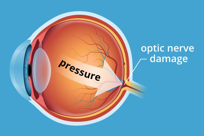
Glaucoma occurs due to a blockage in the drainage system of the eye, due to which the intra ocular fluid gets accumulated leading to the development of high pressure within the eye. This high pressure can damage the sensitive optic nerve, thereby leading to loss of vision.

What are the symptoms of glaucoma?

Slow and often unnoticed decrease of vision and reduced field of vision Headaches accompanied by redness and pain in the eyes Frequent change in glass prescription Reduced vision in low light conditions or at night Seeing coloured haloes or rings around light bulbs

What are the different types of glaucoma?

Primary open-angle glaucoma
It is the most common form of glaucoma where the eye’s natural drainage canals become clogged over time. This is dangerous, because most people have no symptoms or early warning signs. If it is not diagnosed and treated, it can cause a gradual loss of vision.
Open-angle glaucoma treatment
After proper investigations and confirmation of diagnosis, eye drops are used to lower the eye pressure. There are various kinds of eye drops and the doctor will choose the best drop for you depending on associated conditions such as asthma, diabetes, inflammation in the eye etc
Angle-closure glaucoma
It is a type of glaucoma in which the drainage area or angle is closed or narrow. Consequently fluid inside the eyes (aqueous humour) cannot drain out normally which leads to a rise in eye pressure. This may lead to pain in the eye, redness, decreased vision and coloured halos around light. Angle-closure glaucoma may have acute symptoms and need immediate attention in the form of medicines and laser to relieve the acute attack.
Angle closure glaucoma treatment
In addition to drops and tablets, a laser may be recommended to create an alternative opening to relieve the angle closure and allow fluid to pass through and open the blockade (YAG PI). Patients who do not respond to lasers and medicine will need surgery
Secondary glaucoma
Secondary glaucoma develops as a result of other eye conditions such as trauma or injury or complicated surgery, inflammation, use of certain drugs like steroids, etc. Sometimes it may be due to pigment release from the eye (pigmentary glaucoma) or due to deposition of flaky material (pseudo-exfoliative glaucoma). The treatment depends on the cause of glaucoma.
Normal Tension Glaucoma
Also known as low-tension glaucoma or normal pressure glaucoma. In this type, the optic nerve is damaged, despite the pressure remaining normal. Those at high risk for this form include patients with a family history of glaucoma, history of systemic heart diseases, especially rhythm abnormalities.

Glaucoma screening and management at Shakthi Eye Care

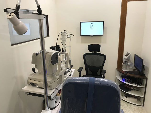
A complete eye examination is needed to diagnose glaucoma. In addition to vision and routine slit lamp examination, eye pressure estimation (Tonometry) followed by evaluation of the drainage angle using a special type of lens (Gonioscopy) is done to know whether a person is having open or closed angle glaucoma. A dilated retinal examination of the retina is important to rule out damage to the nerve. Some of the other tests include visual field testing and scanning of the nerve with the help of specialised equipment (OCT).

What are the modes of treatment available for glaucoma?

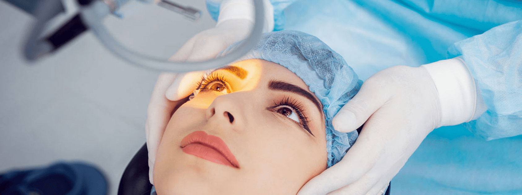
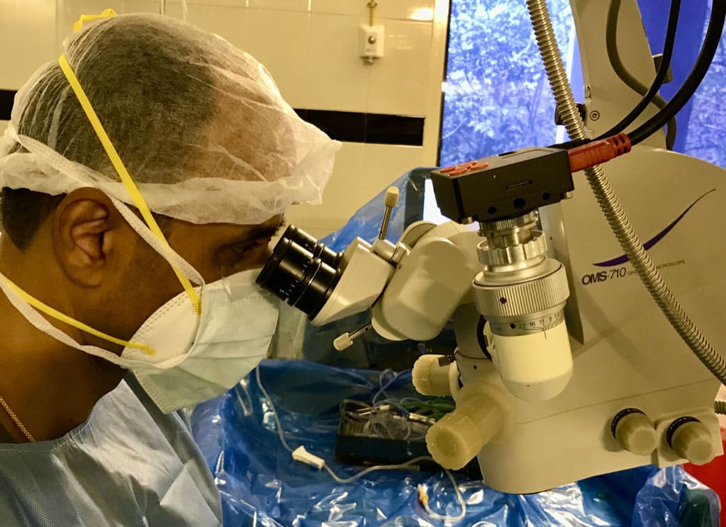
Glaucoma can be treated both medically and surgically.
A number of eyedrops are available that help to reduce the intra ocular pressure. Surgical treatment includes both filtration procedures as well as LASER.

Who is at risk for glaucoma?

  • • People over the age of 40
  • • Family history of glaucoma
  • • Long time steroid users
  • • History of injury to the eye or previous eye surgery
  • • Patients with high power glasses (plus or minus)
  • • Diabetes and hypertension

What is recommended for people with high risk?

People at high risk of glaucoma should get a complete eye exam, including dilated eye examination , once every year. Tests include tonometry, gonioscopy, and in people suspected of having glaucoma, visual field tests to detect glaucoma at an early stage.

eye clinic

Paediatric Eye Diseases

Why screening of children is important?

Screening children at young age is important to avoid childhood blindness which is the second largest cause for blindness in India. Nearly 50% of the causes are preventable and treatable. If not identified and treated at an early age, preferably before 10 years of age, Amblyopia ( lazy eye) sets in which is condition with permanent decrease vision throughout life.

What are the causes of childhood blindness?

Some of the treatable causes of childhood blindness include

Uncorrected refractive errors and squint
Cataract
Glaucoma
Corneal ulcer and scaring
Retinopathy of prematurity-occurs in pre mature babies

When to do vision assessment in children ?

Vision can be tested grossly as early as 6 months after birth and if the baby is suspected of having poor vision in one to both eyes, requires further evaluation to diagnose the cause of poor vision.

Between 3-4 years, vision van be assessed using pediatric symbols (LEA symbols/ HOTV chart).

Four to five year old children can be tested using standard optotypes.

Children should continue to have annual vision examination throughout the childhood and adolescence

What are the common paediatric eye problems which require ophthalmologist consultation ?+

Poor vision for distant objects caused by refractive errors are the most common cause of visual impairment in children and often goes unnoticed by the parents. Symptoms of poor vision include squeezing eyes while watching TV or blackboard, holding things closely to see clearly, poor school performance, frequent complaints of headache and eye strain.

Deviation of eyes in which the eyes look in different directions instead of looking straight (Squint)

Watering with or without redness in one or both eyes

Drooping of eyelid( Ptosis)

Congenital cataract may be seen as a white reflex in the pupillary area and sometimes oscillating eye movements (Nystagmus)

Congenital glaucoma often presents with the child having watering of eyes, intolerance to light and large cornea

eye doctor


opticals shop near me
Glaucoma Awareness Video
opticals
Covid Awareness Video


Blogs

eye clinic in Virugambakkam

Pink Eye

The number of patients with pink or red eyes is on the rise in this season and many people have lots of questions regarding that. This blog is to explain some of the facts related to the red eye.


Read more

Importance of Vision Testing in Children

Many parents are not aware of the fact that it is essential to check vision in all children even if the child is not having any obvious symptoms.


Read more

Contact Lens

The number of people having vision problem due to refractive error is on the rise globally. There are multiple options to improve vision in people having refractive errors.


Read more

Our Gallery

eye clinic in Virugambakkam
hospital in Virugambakkam
Ophthalmologist
SHAKTHI EYE CARE
eye care
eye doctor
SHAKTHI EYE CARE
Dr Balamurugan Sankara Hospital
SHAKTHI EYE CARE
Glaucoma
Cataract surgery
Laser surgery
Cataract surgery

Patients testimonial

See what our treated patients saying..

Doctor is polite and professional. Listens and address the queries properly. Pleasant and peaceful atmosphere

baskar Janarthanam

Very good care and affordable hospital

Sankar Raj


Very good Excellent service. In these days very service minded person. I'll recommend my friends and families to this eye care

BHARATHI


I thank God for getting chance to have the consultation when I needed at the right time. I may give 5 Star due to my limited experience restricted to 4 Stars.

PURUSHOTHAMAN


Very good doctor very knowledgeable and listening our problems very coolly. I can say with one word I.e doctor with professional ethics.

SURESH


Treating very well and atmosphere also very nice

Vijay Kumar


Best hospital, great hospitality and excellent treatment.

Somasundaram


Best eye care centre with affordable consultation fee

Rajesh Kumar Natarajan


I had problems with my eye sight and consulted Dr Balamurugan. He advised me to undergo cataract surgery. He did operation for my cataract and the very next day I can see very well! He is a friendly and very knowledgeable doctor. Many thanks to him and Shakthi Eye Care.

Balamurugan


Hats Off

Vimala


Very Good Service

Thilak


Very genuine

Subhashini


Excellent Service, Doctor so friendly

Mala Murugesan


Excellent service

Sathish


Shakthi Eye Care Centre provides best eye care services in chennai.

Get appointment for treatment.
We follow safety instructions.

Book an Appointment

eye hospital near me

Shakthi Eye Care Centre

  • Shakthi Eye Care Centre, No.9/73, Elango Nagar Annexe, Virugambakkam,
    Chennai - 600 092.
  • (+91) 044-23774676| (+91) 63694 70626
  • shakthieye@gmail.com
  • www.shakthieyecare.com
Opening Hours
  • Monday - Saturday :
    6.00 pm - 9.00 pm
  • Sunday :
    Closed

Connect With Us

Copyright ©2025 Shakthi Eye Care. All Rights Reserved | Designed by Tryambaka Techno Solutions | Privacy Policy